The X-ray beam from the tube-head should meet the tooth and the image receptor at right angles in both the vertical and horizontal planes. However the anatomy of the oral cavity makes it.

Periapical Radiography Pocket Dentistry
Fluoride given to children and adults after a prophy.

. Panoramic x-ray of full mouth. The patients oral hygiene. It is possible for a foreign body to enter the airways and cause choking.
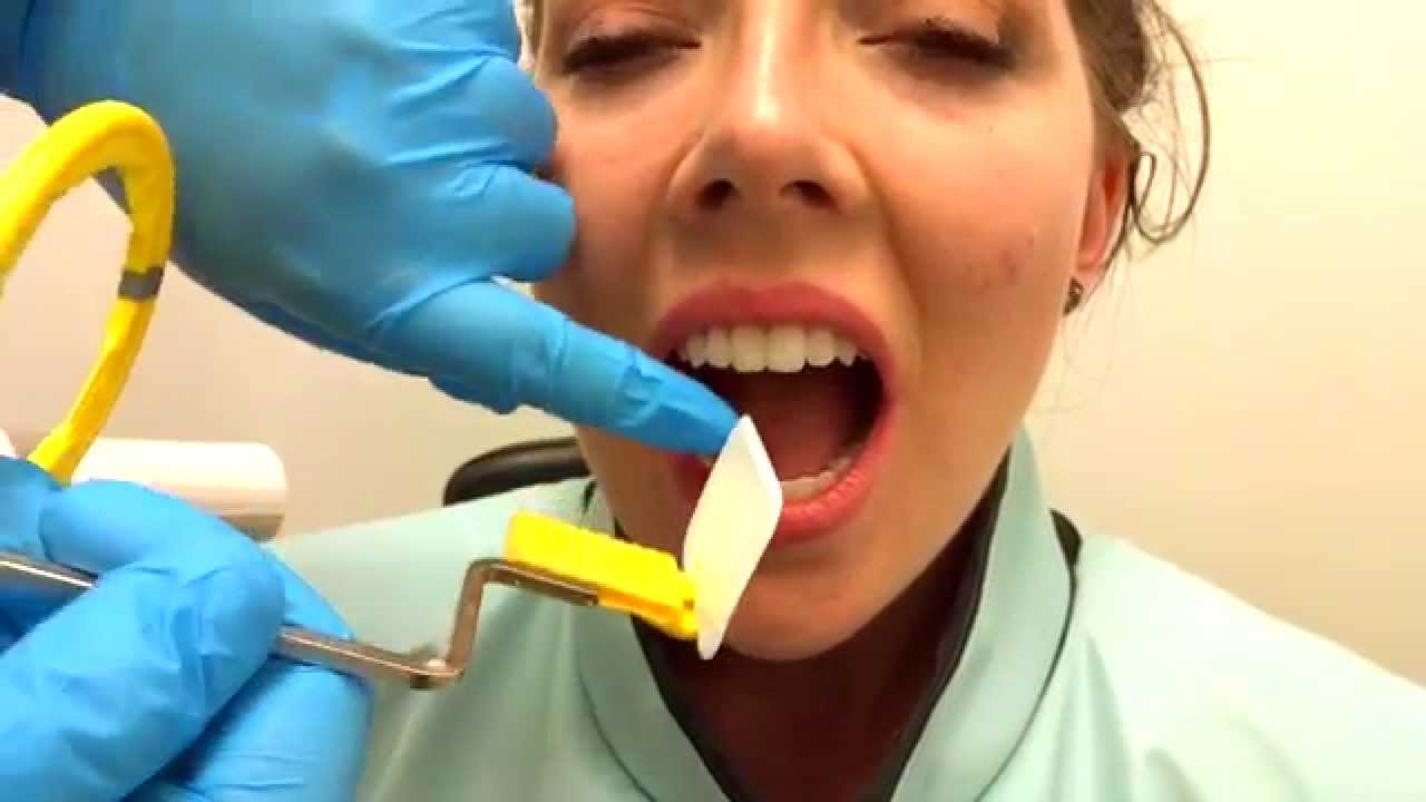
In one study peanuts were the most common obstruction. Periapical x-ray of one tooth. Positioning should be reproducible.
Some dental practices have a separate room for X-rays while others perform them in the same room as cleanings. Intra-oral radiographic techniques. A choking case can require the fast usage of basic anti-choking techniques to clear the airway.
The X-ray machine is positioned alongside your head to record images of your mouth. The tooth under investigation and image receptor should be in contact or as close together as possible. Lateral X ray showing a 9mm battery in the intestines Multiple button batteries in the stomach Airways.
20-gauge needle usually used for upper-tooth injections. 24-gauge needle usually used for lower tooth injections.

Periapical Radiography Pocket Dentistry

Periapical Radiography Pocket Dentistry
How To Take Periapical Radiographs Youtube

Periapical Radiography Pocket Dentistry

Periapical Radiography Pocket Dentistry

How Make Periapical X Ray

Periapical Radiography Pocket Dentistry
Periapical Radiography Pocket Dentistry
0 komentar
Posting Komentar